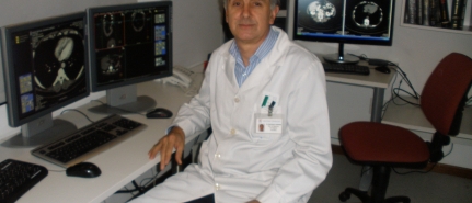
El HOSPITAL BRITÁNICO ALCANZA LA DIGITALIZACIÓN TOTAL DE SU DEPARTAMENTO DE DIAGNÓSTICO POR IMÁGENES En el marco de su política de mejora continua, el Hospital Británico alcanzará en los próximos días la digitalización del cien por ciento de las imágenes obtenidas en los distintos procedimientos y estudios realizados por su Departamento de Diagnóstico por Imágenes. De la mano del avance tecnológico, los viejos departamentos de Radiografía fueron evolucionando hasta llegar en nuestros días a ser departamentos o áreas de Diagnóstico por Imágenes, donde importa tanto o más que la tecnología la gestión del propio equipamiento así como de las imágenes que se obtienen mediante los distintos estudios y procedimientos que se realizan. El Hospital Británico cuenta con un equipamiento de última generación que gestiona conforme estándares de calidad mundial, que se traducen en una mejor atención a los usuarios, ya que ayuda al diagnóstico y la terapeutica, y brindan seguridad total en la conservación de la información. Con el comienzo en los próximos días de la digitalización de las imágenes obtenidas en los estudios de mamografía, el hospital alcanzará el cien por ciento de almacenamiento digital de las imágenes obtenidas en estudios y procedimientos realizados por su Departamento de Diágnostico por Imágenes. Así, las mamografías se sumarán a radiografías, ecografías, resonancias, tomografías, angiografías y demás estudios imagenológicos que se realizan en el hospital, cuyos resultados se registran y almacenan digitalmente. La digitalización de las imágenes obtenidas con estos procedimientos representa un gran potencial en términos de accesibilidad y seguridad. Así, por ejemplo, imágenes e informes pueden ser consultados por el médico desde puntos remotos como piso, block quirúrgico, consultorio o emergencia, y por dos o más profesionales al mismo tiempo. Las imágenes, así como los informes médicos correspondientes, se integran asimismo a la historia clínica electrónica, con lo cual toda la información de cada paciente se almacena y respalda de forma digital. El Departamento de Diagnóstico por Imágenes del Hospital Británico atiende a más de 7.000 usuarios por mes, señaló el doctor Nelson Di Trápani, Jefe Médico del departamento. Las imágenes obtenidas son registradas digitalmente con un código que, a su vez, las relaciona con el informe médico correspondiente. La calidad de la imagen y la posibilidad de visualizarlas en 3D presenta un gran potencial para los médicos que pueden ampliar, girar, medir, comparar imágenes, analizar estructuras y apelar a otras herramientas a las que de forma convencional no accedían, explicó. Para capturar y almacenar las imágenes se utiliza un formato de calidad médica de imágenes denominado DICOM 3, que es un estándar mundial, y a su vez se utiliza un sistema llamado PACS, que es otro estándar para el manejo de las imágenes, sostuvo Di Trápani.